Hi everyone,
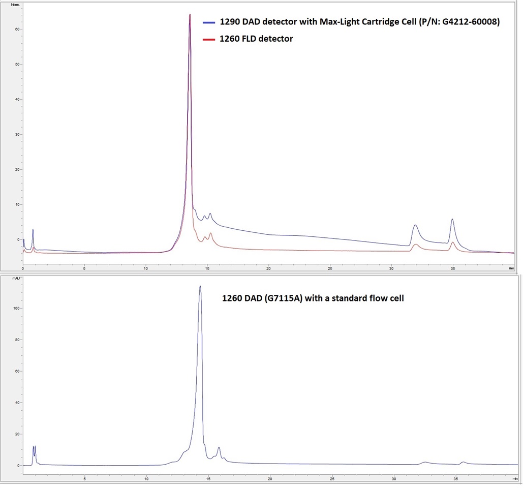
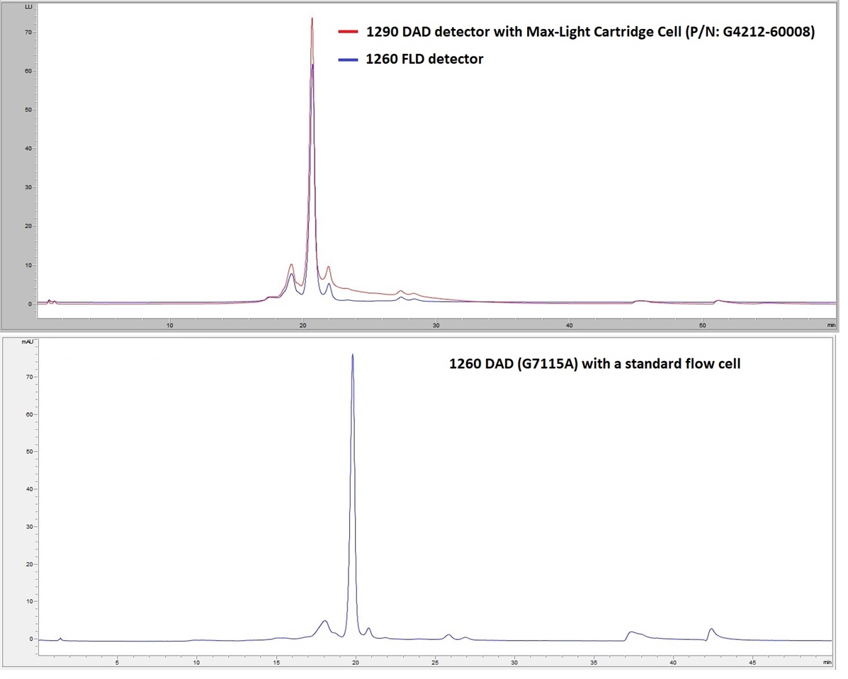
I have got a problem with 1290 DAD detectors in salt gradient ion exchange chromatography of proteins, I have been developing different salt gradient ion exchange methods for the charge variants analysis of monoclonal antibodies, for most of the methods, I observe a kind of peak tailing or baseline drift when using 1290 DAD detectors with Max-Light Cartridge Cell (P/N: G4212-60008), but not in the case of 1260 DAD (G7115) with a standard flow cell (G1315-60022) and also a FLD detector, I have two examples as follows:
Example 01:
Sample: Monoclonal antibody A
Mobile Phase A: 20 mM NaH2PO4, pH= 6.2
Mobile Phase B: 20 mM NaH2PO4, 200 mM NaCl, pH= 6.2
HPLC column: TOSOH CM-STAT (4.6×100 mm), P/N: 0021966
5 minutes with 5% of the mobile phase B then a gradient from 5 to 50% of mobile phase B in 25 minutes.
Detection wavelength with DAD: 280 nm, bandwidth: 4 nm (reference wavelength: 360 nm, reference bandwidth: 100 nm);
FLD detector: Excitation 280 nm, Emission 340 nm
I have used the DAD and FLD detectors in series for the chromatogram in the top.

Example 02:
Sample: Monoclonal antibody B
Mobile Phase A: 10 mM NaH2PO4, pH= 6.4
Mobile Phase B: 10 mM NaH2PO4, 200 mM NaCl, pH= 6.4
HPLC column: ProPac WCX-10 (4×250 mm), Thermo, P/N: 054993
5 minutes with 100% of the mobile phase A then a gradient from 0 to 25% of mobile phase B in 38 minutes.
Detection wavelength with DAD: 280 nm, bandwidth: 4 nm (reference wavelength: 360 nm, reference bandwidth: 100 nm);
FLD detector: Excitation 280 nm, Emission 340 nm
I have used the DAD and FLD detectors in series for the chromatogram in the top.

I have tried different instruments, different reference wavelengths and also a Bio-inert Max-Light Cartridge Cell with no significant improvement in the peak shape and baseline.
I appreciate if anyone could share their experience to solve this probelm.
Regards,
